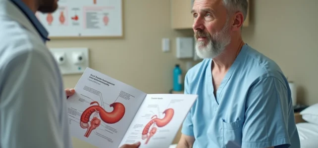
An external hemorrhoid is a venous dilation located under the skin of the anal margin, visible to the naked eye without medical instruments. Recognizing this lesion in a photo requires the ability to distinguish a simple skin swelling from an acute thrombosis, and especially not to confuse a hemorrhoid with a more serious anal pathology. Identifying the correct visual criteria helps guide the approach but never replaces a clinical examination.
Anatomy of the anal margin and precise location of external hemorrhoids
Hemorrhoids are not anomalies. They are vascular structures present in everyone since birth, composed of blood lakes associated with small arterial and venous vessels. They are distributed in three to four cluster-like groups, fixed to the wall of the anal canal.
Recommended read : How to find a phone number using an effective and free reverse directory
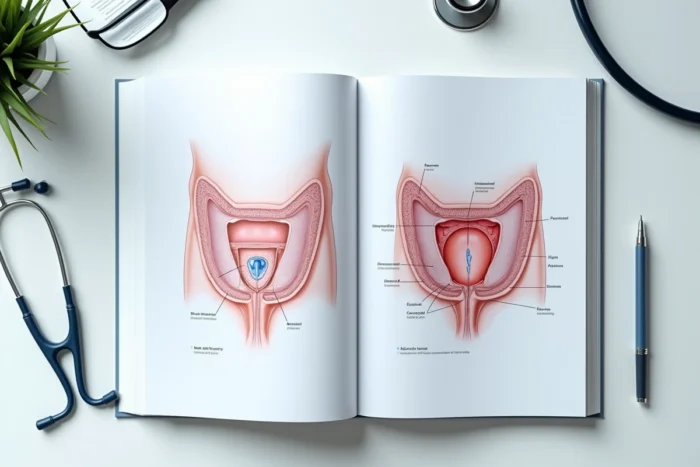
The anatomical distinction between internal and external hemorrhoids is based on their position relative to the pectinate line of the anal canal. Internal hemorrhoids line the inside of the canal and are only visible with an anoscope. External hemorrhoids, on the other hand, are located just beneath the skin that borders the anal opening.
In a reliable photo, this location is represented by a swelling covered by skin (and not mucosa), often bluish or purplish. The skin covering the external hemorrhoid is innervated, which explains the pain upon touch, unlike internal hemorrhoids which remain painless for a long time. To see photos of external hemorrhoids on Doctinews, one must keep in mind this criterion of subcutaneous localization to accurately interpret what is observed.
You may also like : How to Easily Buy and Sell Your Car Using Online Solutions
External hemorrhoidal thrombosis: what a reliable photo really shows
The majority of image searches actually concern external hemorrhoidal thrombosis, the most dramatic and painful form. This complication occurs when a blood clot forms in an external hemorrhoidal vein.
In a quality photograph, thrombosis can be recognized by several precise visual criteria:
- A well-defined, firm nodule upon touch, located at the anal margin and not inside the canal
- A characteristic bluish-purple coloration, related to the stagnation of coagulated blood under the skin
- A tense appearance of the overlying skin, sometimes shiny, indicating edema and pressure from the clot
- A variable size, ranging from a pea to a marble, appearing suddenly within a few hours
The appearance is typically sudden. The pain is immediate, sometimes intense, and worsens when sitting or during defecation. This rapid chronology distinguishes thrombosis from a simple progressive inflammatory swelling.
Visible evolution in photos at different stages
An untreated thrombosis evolves spontaneously over a few days. The bluish swelling gradually loses its tension, the pain decreases, and the clot partially resolves. A residual tag, a small fold of flaccid skin at the anal margin, may persist, sometimes mistakenly confused with an active hemorrhoid in photos.
Differentiating a tag from an ongoing thrombosis is a point often misunderstood. The tag is soft, painless, flesh-colored. The thrombosis is firm, painful, and bluish. A photo taken after partial regression may show a misleading intermediate appearance.
Risks of confusion with other anal pathologies in photos
This is the most underestimated point in online content about hemorrhoids. The visual symptoms (swelling, redness, minor bleeding) overlap with those of other serious anal pathologies: anal fissure, perianal abscess, and in some cases, anal or colorectal cancer.
A perianal abscess produces a red, hot, painful swelling that can resemble hemorrhoidal thrombosis in a poorly framed or low-resolution photo. The difference lies in the associated fever, fluctuation upon palpation, and rapid progression to suppuration, elements impossible to evaluate from an image.
An anal fissure causes pain and bleeding similar to that of a hemorrhoid, but the lesion is a linear wound of the anal canal, rarely visible in a photo taken by the patient themselves.
The trap of self-diagnosis by image
Attributing anal bleeding to hemorrhoids without medical examination sometimes delays the diagnosis of more serious lesions. Campaigns for colorectal cancer prevention increasingly warn against this drift. Bright red bleeding on toilet paper often falsely reassures, especially if the person has already visually identified an external hemorrhoid.
The rule is simple: any persistent anal bleeding or any pain that does not subside within a few days warrants a consultation, regardless of what a photo found online suggests.

Visible triggering factors in photos of acute crises
External hemorrhoids evolve in crises. Photos taken during an acute phase show significantly more pronounced swellings than those taken during a calm period. Understanding the triggering mechanical factors helps to contextualize what is observed.
Repeated straining during defecation remains the main factor. Chronic constipation, frequent episodes of diarrhea, and carrying heavy loads increase venous pressure in the external hemorrhoidal plexus, promoting the formation of thromboses.
In crisis photos, perianal edema often accompanies the thrombosis itself, giving a more diffuse swollen appearance than the simple isolated nodule. This presentation may cause more concern, but it corresponds to the same pathology at a more pronounced inflammatory stage.
Criteria for reliability of an online photo of an external hemorrhoid
Not all images found on the internet are equal. A useful photo for identification must meet several conditions:
- Come from an identifiable medical source (scientific society, hospital site, named dermatologist or proctologist)
- Include a caption specifying the stage and type of lesion photographed
- Show sufficient framing to locate the lesion relative to the anal margin
- Not be retouched or compressed to the point of altering the actual colors of the skin
Photos from generic image banks or forums without medical moderation carry a high risk of misidentification. An image without a clinical caption has no diagnostic value.
The French National Society of Coloproctology publishes annotated images by practitioners, with photographic credit. This type of source remains the reference for comparing one’s own observations to documented cases, without falling into the traps of approximate self-diagnosis.